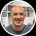
Exclusive Interview with Dr. Jason Hodges
I am extremely fortunate to not only have a loyal group of newsletter subscribers, but also a very knowledgeable and passionate group of individuals who come from unique backgrounds. Collectively, you subscribers give me a ton of outstanding feedback that makes me better at what I do.
After Newsletter 95, I received a great email response from Dr. Jason Hodges:
Regarding the low back, I am a radiologist and I see MRIs every day describing what you said in the newsletter. Lots of people have bulging discs without symptoms. This is especially true of older patients who can have bulging discs at every level but without focal neurologic symptoms. In my experience, younger patients tend to have focal neurological signs with even mild disc bulges or disc herniations. But very often, the symptoms don’t match up with the imaging findings. I have seen patients with symptoms down the right leg, but the disc herniation is on the left side, etc.
Needless to say, that “etc.” at the end of the last sentence got me intrigued, so I asked Dr. Hodges if he would be willing to do an interview for our subscribers. I think you’ll find it very enlightening – and forward-thinking.
EC: Thanks for joining us this week, Dr. Hodges. Could you please tell us a bit about both your professional background and health and human performance interests?
JH: Thanks for the opportunity, Eric. I did my undergrad at University of Kansas with a BA in biochemistry graduating in 1991, and received my MD degree from U. of Kansas School of Medicine in 1995. I finished my Radiology residency at U. of Missouri in 1999 and received my American Board of Radiology certification the same year. I am currently an executive partner in S and D Medical LLP in NYC. My interest in fitness really lies outside my professional duties although there is obviously some overlap. My Radiology training is not specific to fitness.
EC: In your reply to my newsletter last week, you not only confirmed some of the things I noted about MRI results in what we think are healthy lower backs, but also had some other very interesting experiences to share. Would you please fill our readers in?
JH: Often imaging findings do not correlate with clinical findings. Older patients often have very degenerative spines without symptoms. Whereas younger patients can have small bulging discs or herniated discs and have debilitating pain. The human body has a great reserve capacity. I see many “normal” kidneys that are in chronic renal failure
Medical imaging generally deals with anatomy: how organs “look”, not so much how they function. Obviously, they are linked, but function can decline long before anatomic changes occur. Symptoms can occur without imaging abnormalities. This leads doctors to conclude that nothing is wrong because the x-ray/CT scan/MRI looks normal. This is simply not the case.
Medical imaging is simply one piece of the clinical puzzle. An analogy can be made with astronomy. You can image the universe at visible light, x-ray, ultraviolet, infrared, etc. Each modality provides a vital, but incomplete picture of the universe. You have to put it all together to get the big picture.
EC: How about the knees? I know a lot of people are walking around with chronic ACL tears that aren’t symptomatic, but what else do you see?
JH: It is often easier to see acute injuries better than chronic images. We often see the secondary finding, such as edema or fluid collections rather than the direct injury itself. An acute ACL tear may show a gap in or fraying of the ACL, surrounding edema and joint effusion. A chronic ACL tear may show only a wavy appearance or abnormal signal as scar tissue has partially healed the injury. But it is important to recognize the chronic ACL tear because it alters the biomechanics of the knee, stressing other parts of the knee. This can lead to a higher risk of meniscal tear or premature arthritis. A common cluster of findings in acute knee injury is ACL tear, medial meniscal tear and medial collateral ligament sprain/tear and a joint effusion.
EC: Shoulders?
JH: The most common finding I see is tendinopathy of the supraspinatus tendon. It is the most likely to be impinged under the acromion and clavicle. The shape of the acromial hook can predispose to impingement, as can arthritic changes of the acromioclavicular joint. In radiology, we tend to use the term “tendinopathy” rather than “tendonitis”. “Tendonitis” implies white blood cell inflammation, which we cannot confirm on MRI. So we use the imaging term of “tendinopathy” which can certainly include tendonitis.
EC: So, what’s your take? Are we too heavily reliant on MRIs as a society? Certainly, it takes a lot more resources to get a MRI than x-rays, yet many people seem to request these at a moment’s notice to gain some peace of mind. What kind of accuracy are we talking?
JH: As I said, MRI is just a piece of the big picture. Some of the limitations include the fact that we image the joints in a static state, in one position. We image the lumbar spine with the patient lying down which is a whole different loading scheme than standing up. The tracking of the patella during extension is really best assessed by physical exam, not by MRI. It is a matter of putting too many eggs in the imaging basket, so to speak. MRI is the best imaging modality for the soft tissues, but it is not all-seeing/all-knowing.
EC: Let’s talk about lifters. What are you seeing in terms of chronic adaptations to lifting heavy stuff?
JH: To be honest, we don’t image many lifters except in the setting of acute injury. Lifters tend to be younger and healthier. Certainly, lifters have better bone density and have a lower risk of osteoporosis. Larger muscles and lower bodyfat are obviously the case.
EC: Aside from lifting, what other lifestyle habits have you found lead to less-than-stellar diagnostic imaging? Alcohol? Certain occupations?
JH: By far, the biggest limitation is obesity. All of the imaging modalities are limited by it, mostly for technical reasons. An ultrasound beam can only penetrate so far into the soft tissues. X-rays and CT scans are degraded by scattered radiation, which leads to a higher radiation dose and grainy images. Also, the time it takes to do the study increases, which gives a higher incidence of motion blur.
EC: So diagnostic imaging is less accurate with obese patients? One more reason to not get fat in the first place!
We often talk about how the best doctors are the ones who meet the lay population halfway. In other words, they’re the ones who can tell an injured patient what he CAN do, and not just what he CAN’T do. My experience has been that the best trainers and coaches are the ones that can meet the doctors halfway, and it’s something to which I attribute a lot of my success.
To that end, what resources would you recommend to trainers, coaches, and everyday weekend warriors looking to learn more in the direction of the clinical realm?
JH: Frankly, the mainstream media is not a great source of information. It is incumbent on us radiologists to let the primary care doctors know what we can image and, more importantly, what we can’t. Orthopedic surgeons tend to be the most knowledgeable regarding the musculoskeletal system, but don’t discount chiropractors. I am pretty open-minded to alternative medicine, unlike many of my fellow MDs. My chiropractor does a great job using ART on my trigger points in my traps. Also, never be afraid to get a second opinion.
My advice to all practitioners – be they doctors, chiropractors or trainers – is to learn as much as possible. Be confident in your knowledge and abilities, but don’t think that any one practitioner has all the answers. Medical knowledge is too vast for anyone to know everything about everything. I know my medical school training regarding fitness and nutrition was paltry. Sure, I learned about muscle fiber composition and the biochemistry of vitamins and minerals. But, most doctors just parrot the standard dogma of low-fat, high-carb diet, walk 20 minutes three times a week, etc. You and I both know that won’t lead to any significant body composition changes.
EC: Agreed. I actually know several doctors who have “seen the light” when they’ve started to read more of Dr. John Berardi’s work – not to mention the latest research of carbohydrate-restricted diets from the likes of Jeff Volek and Cassandra Forsythe. What else?
JH: Seek out those practitioners who aren’t afraid of the cutting edge. Just as you wouldn’t want a trainer who is a glorified rep counter, you don’t want a doctor who is simply going to give you the same old tired, old-school nutrition and fitness “advice,” if you can even call it that. That advice may promote health, but it won’t give the body composition changes most of your readers seek.
In addition to being confident in their abilities, practitioners need to know when to refer to other people. Some things need to be treated medically or surgically. They can’t be fixed in the gym or at the training table. Health and wellness should be a team effort with everybody working in their areas of expertise and not outside of it. Underconfidence and overconfidence in your abilities are equally bad for your client/patient.
Unfortunately, Western style medicine is very disease-oriented and body-part-oriented, often losing the big picture, especially regarding the whole kinetic chain of the musculoskeletal system. My opinion is that this is where trainers and chiropractors shine. I wish my fellow doctors would be more amenable to referring patients to trainers/chiropractors for problems that don’t need medical or surgical treatment. The human body has great ability to adapt and heal itself, if you give it a chance.
EC: Thanks again for taking the time to be with us!
JH: My pleasure. Thank you for inviting me. My kind gratitude to my colleague D. Dillon, RN, BSN for her assistance.


