Thinking Beyond Diagnostic Imaging
About ten years ago, I was in the operating room to observe my first Tommy John surgery. Much like my time in gross anatomy class during my undergraduate studies, it was an invaluable experience that helped me to appreciate human structure (and, in turn, movement) in a way that anatomy textbooks couldn’t offer.
Textbooks typically present a very “neat” anatomy where muscles, tendons, ligaments, bones, and nerves are predictably positioned. You can imagine my surprise, then, when the surgeon made the initial incision along the medial elbow and it yielded a bunch of “stuff” in the way. The fascia, the intermuscular septum, the ulnar nerve, and a host of other unrecognizable structures make you realize that a) every anatomy course or textbook you’ve ever undertaken hasn’t done justice to what’s really going on at the elbow (or anywhere else in the body, for that matter), b) it takes a lot of practice to become a great surgeon, and c) you shouldn’t let just anyone cut you open for surgery.
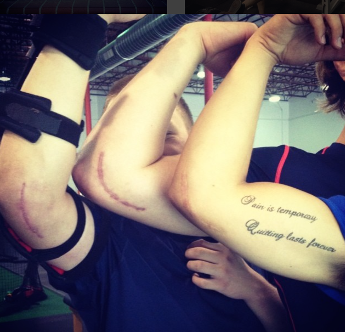
Now, let’s fast-forward to the post-operative timeline. After the repair is complete and the patient is stitched up, the elbow is splinted at 90 degrees of flexion for a week. Following that week, the arm is put in a hinged brace that gradually allows more motion over the course of weeks 2-6. After six weeks, the brace goes away. In short, it’s quite a bit of time with the elbow in a limited range-of-motion situation as a means of protecting the repair.
Not surprisingly, some patients have a lot of trouble getting back their motion – both from the graft gradually stretching out and the musculotendinous structures regaining their length. We’d be crazy to think that the aforementioned fascia structures aren’t implicated in the challenges of regaining ROM, though. And, if they’ve got a role in limiting ROM, they’ve certainly got a role in the associated stiffness (and, sometimes, pain) that post-operative patients feel. Here’s where a variety of manual therapy interventions – ranging from dry needling to instrument-assisted work – have yielded outstanding results. While some folks like to scream and shout in opposition to this fact, it’s hard to refute that manual therapy has endured the test of time to the tune of thousands of years.
Massage has been around for 4,300+ years, yet there are still people out there who deny the value of manual therapy? pic.twitter.com/uMUSPpPpJK
— Eric Cressey (@EricCressey) May 24, 2016
Only recently have there been technology advancements that allow us to better understand the role of the fascia system with respect to pain and performance (Bill Parisi spoke to this on our podcast a while back; listen here). In a seminar not too long ago, my friend Sue Falsone highlighted some great evidence on the “decompression” that takes place in this regard with a cupping intervention.
Now, let’s take a step back and think about the big picture of diagnostic imaging. When we have chronic pain that alters movement patterns, we have adaptive changes of the fascial system. While the diagnostic imaging – MRI, CT scans, x-rays – might pick up on the structural defect, it might overlook the compensatory changes to the fascial system (much of which overlays the actual injury) that can’t be appreciated by these types of scans.
When we look at chronic shoulder pain, is the problem only the rotator cuff tear? Or is that problem magnified by the fascia limitations that arise from years of avoiding various ranges of motion (including at adjacent joints) that would normally be accessible?
I’ve written extensively about having both a Medical and Movement Diagnosis. The truth is that the discussion of the fascia system is probably a happy medium between the two. We may think too much about the injury the diagnostic imaging identifies and too little about the overlying and surrounding tissues. At the other end of the spectrum, we may be too quick to define a movement limitation as joint, musculotendinous, ligamentous, or motor control without first considering the role the fascial system is playing.
What’s the most important lesson here? Professionals from all walks of sports medicine need to work together to thoroughly evaluate injuries and movement competencies in order to design the best performance and rehabilitation programs. And, we need to remain openminded to new technology that may make it easier for us to take an even more accurate and individualized approach to each case.
In this vein, I’d highly recommend checking out my Thoracic Outlet Syndrome course, as this challenging diagnosis is a great example of how a condition often can slip past common diagnostic imaging.


